


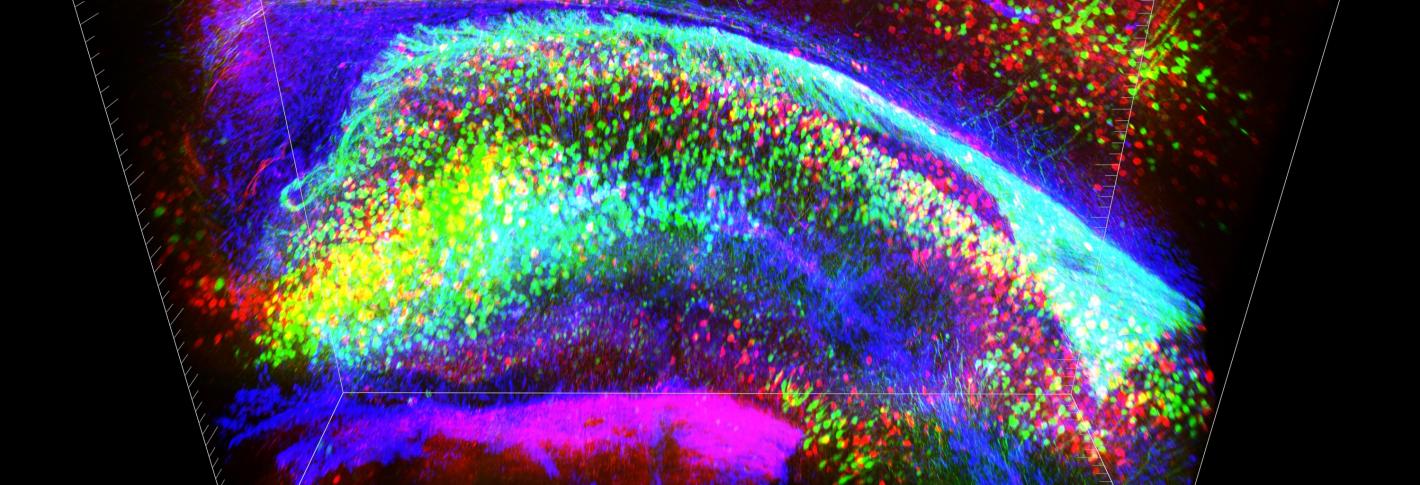
To understand role of neurons and the circuits in which they participate neuroscientists must be able to gather data on a neuron’s electrical activity, such as when they fire, in real-time. Picower scientists are constantly innovating new genetic and chemical sensors, as well as electronic and imaging-based means to track neural activity both in vitro and in vivo and develop sophisticated means to analyze the large volumes of data gathered.
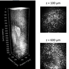
In many ways, Picower Institute neuroscientists are explorers for whom new ways to see inside the brain are essential for finding answers to their questions about how the brain works at scales ranging from synapses to whole networks. Researchers at the institute doesn’t just apply the latest imaging techniques, it often creates new technologies to make imaging better.
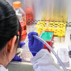
Biological research often calls for imbuing cells, tissue, or animal models in the lab with specific new capabilities – or disabilities, for instance to observe the differences between altered and unaltered cells. Picower Institute neuroscientists employ advanced techniques such as CRISPR/Cas9, stem cell, and transgenics to conduct such experiments.
By engineering cells with light-responsive ion channels, optogenetics allow the activity of cells such as neurons to become controlled by pulses of visible light. The technology is widely used throughout the institute in experiments in which purposeful instigation or suppression of neural activity can reveal important data on the functions of cells, circuits, systems, and behaviors.
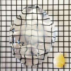
To best visualize and study neural tissue, sometimes researchers need to make chemical alterations, for instance to clear away opaque lipids or to label different molecules. Innovative technologies such as CLARITY, MAP, stochastic electrotransport and SWITCH that can make neural tissue optically clear, enlargeable to allow neuroscientists to better image and study the structure of the brain.