

September 29, 2026
When you have a sore throat, your doctor can take a swab and culture it to determine if it’s strep. When you see something weird on your skin, a dermatologist can take a snip for a biopsy. In both cases, the providers will typically be able to come back to you with an objective, biologically based indication about what’s going on with your health and use that to guide your care.
But the brain cannot be swabbed or biopsied. For a long time, psychiatrists and neurologists have had to rely solely on behavioral symptoms, which can be frustratingly subjective and imprecise. In 2010, for instance, Dr. Thomas Insel, then director of the National Institute of Mental Health, called for psychiatry to move from relying solely on symptoms to a basis in genomics and neuroscience to benefit from the same kinds of biomarkers other medical practitioners can employ.
Developing informative, minimally invasive biomarkers of brain health has required years of fundamental research. Scientists have made headway—for instance by associating certain genetic variations with diseases or disease risk—and indeed a lot of progress is evident in Picower Institute research. Including in some of their most recent studies and current projects, Picower Institute researchers have been developing potential biomarkers for autism spectrum disorders, Alzheimer’s disease, bipolar disorder, and even coma and unconsciousness under general anesthesia. Biomarkers not only provide an objective, physical basis for diagnosis, they can also make treatment more personalized and track treatment progress.
Take the case of bipolar disorder. Because patients exhibit depressive episodes and then sometimes manic ones, they might at first be diagnosed with depression and given related medications which may have side effects, said William R. and Linda R. Young Professor Elly Nedivi. If they first present to a clinician as manic, that could be confused with schizophrenia. And if they see their doctor as a child, either the manic or depressive state might present differently than it would if they were adults. The conflation of age-related changes, potential misdiagnosis, and misdirected medication side effects can all combine to make a correct bipolar diagnosis hard to achieve from snapshots of behavioral observations alone. Except that’s pretty much all that’s currently available.
“Diagnostics are very complex and really prone to not being correct,” Nedivi said. “There is no objective marker.”
Genes and risk
To help the 2 percent of the population who experience bipolar disorder, Nedivi’s lab has been laying the groundwork to develop genetic biomarkers for bipolar disorder risk. Her lab has shown that specific mutations in a gene called SYNE1 that have been significantly associated with risk for bipolar disorder undermine the expression of a protein called CPG2, potentially affecting communication between neurons. The data her lab has been developing raise the possibility that when a future patient presents to their clinician as either depressed or manic, the doctor will be able to administer a genetic screen that will flag the risk-imparting mutations. This would provide a powerful, objective signal that the real underlying issue might be bipolar disorder, possibly prompting the patient’s care providers to consider that diagnosis when they wouldn’t have otherwise.
Another disorder where biomarkers are sorely needed is Alzheimer’s. While few good treatment options exist, the degenerative nature of the disease means that whatever interventions are tried, they’ll be more helpful if they are delivered as early as possible, said Picower Professor Li-Huei Tsai.
“Biomarkers are accessible and quantifiable,” Tsai said. “Most importantly, they can serve to help early diagnosis—before the appearance of symptoms—for prevention measures.”
Over the last few decades, researchers have identified many Alzheimer’s risk genes. For instance, the largest, the APOE4 variant of the APOE gene, confers a 3-fold risk of developing the disease on people with one allele and a 10-fold risk on those who carry it on both their copies of chromosome 19.
Tsai’s lab has invested heavily in understanding how APOE4 and another genetic risk variant, ABCA7, contribute to Alzheimer's risk. In several studies over the last several years, her lab has shown that the variants contribute to problematic lipid dysregulation in various cell types in the brain (see our Winter 2025 edition). In a project funded by the Cure Alzheimer’s Fund, Tsai’s lab is investigating whether lipid levels and composition in cerebrospinal fluid (CSF) change with age, sex, APOE variant, and whether someone has Alzheimer’s. Significant changes in CSF lipids could act as accessible biomarkers of disease progression that account for patient sex and APOE variant.
Tsai’s studies of APOE4 and ABCA7 each pointed to a particular nutrient, choline, as a potential treatment to restore normal lipid regulation, at least in some cell types. While one can increase choline by eating eggs, meat, fish, and some beans or nuts, Tsai and colleagues at the University of Texas and MD Anderson Cancer Center have been collaborating to run an early-stage clinical trial testing high-dose choline supplements for six months with APOE4 carriers. One of the first things they are looking for are potential biomarkers accessible through CSF or plasma that would indicate if the nutrient is affecting factors that contribute to Alzheimer’s risk.
Tsai is also developing potential Alzheimer’s biomarkers in two other research collaborations. FINGERPRINT is an AI platform trained on many different Alzheimer’s datasets (e.g. genomic, proteomic, clinical assessments and lifestyle indicators) to produce new integrated, personalized biomarkers that might help predict Alzheimer’s risk. And in a new project funded by the Massachusetts Life Sciences Center, Tsai and MIT Computer Science Professor Manolis Kellis are investigating possible biomarkers that can help women better assess cognitive risks from hormone replacement therapy.
While Alzheimer’s and bipolar disorder are examples of disorders where many genes might contribute to an overall risk, some other conditions have well known single-gene causes. Even then, research is showing, it can be important to go beyond the assumption that dysfunction of the gene is an all-or-nothing problem.
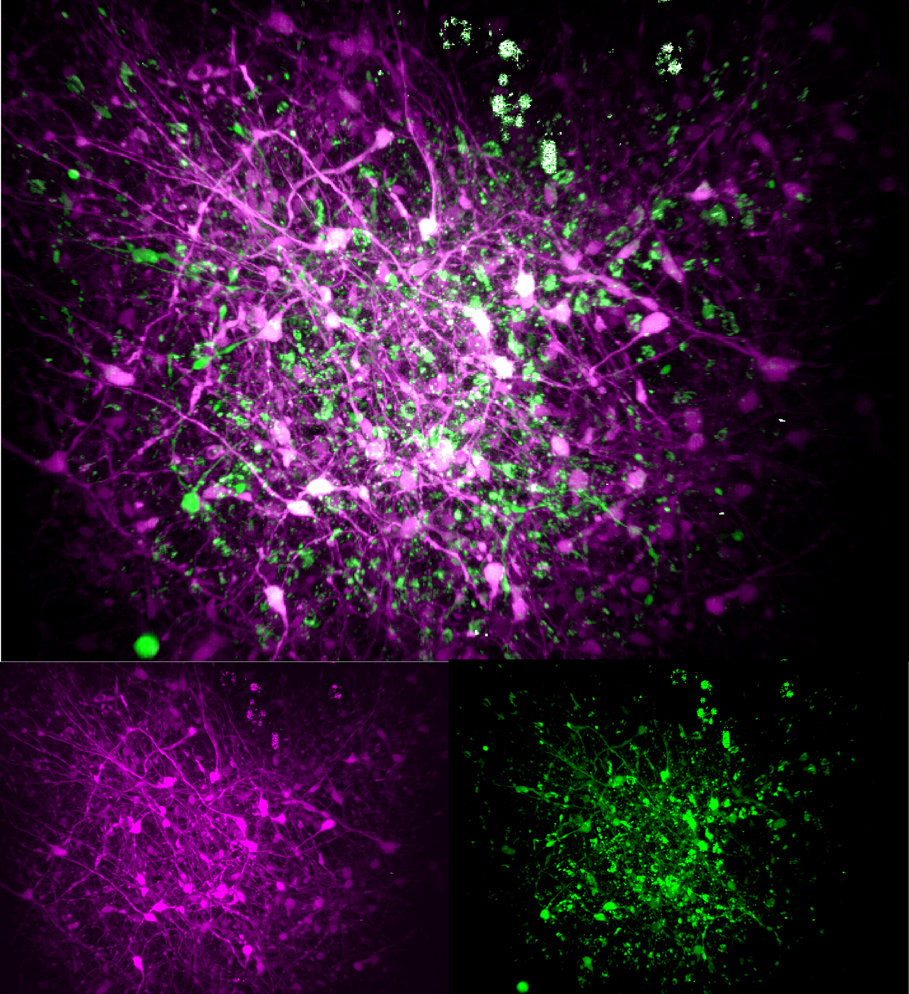
For instance, Rett syndrome is a severe autism-like neurodevelopmental disorder arising from mutations that impede the function of a gene called MECP2. Many lab studies of Rett syndrome, for instance in mouse models or using tissue cultures, simply disable the gene wholesale. But in a study in Nature Communications earlier this year, the lab of Newton Professor Mriganka Sur, led by postdoc Tatsuya Osaki, used advanced human “organoid” tissue cultures that blend multiple cell types to investigate the consequences of two different MECP2 mutations. Importantly, they found that each mutation affected the organoids in distinct ways and that rescuing the specific deficits required different interventions. Although there is an overall treatment for Rett syndrome (developed based on Sur’s research), the results suggest that a patient with one Rett-related mutation might also benefit from different treatments than a patient with another one.
Brain Waves
Like Rett syndrome, fragile X syndrome is a neurodevelopmental disorder that arises from mutation and loss of function of a specific gene: FMR1. In fact, it is the most common inherited form of autism. While the gene itself is a biomarker of the disease, it of course does not change during a patient’s lifetime. So what’s needed is a readily measurable biomarker that could help clinicians track the progress of potential therapies, complementing behavioral assessments. Earlier this year in Nature Communications, the lab of Picower Professor Mark Bear led by postdoc Sara Kornfeld-Sylla revealed a specific pattern of brain waves, measurable with EEG electrodes on the scalp, that could provide just such a biomarker.
In their study, Bear and Kornfeld-Sylla’s team measured the brain waves of human boys and men, with or without fragile X syndrome, and comparably aged male mice, with or without the genetic alteration that models the disorder. Using a novel analysis approach, Kornfeld-Sylla uncovered specific and robust patterns of differences in low-frequency brain waves between typical and fragile X brains shared between the species at each age range. Moreover, in the mice they showed that the biomarker was able to indicate the effects of even single doses of a candidate treatment for fragile X called arbaclofen.
It’s especially exciting that the brain wave biomarker proved to be consistent between mouse models and human patients, Bear said. It raises the possibility that scientists could use it to assess whether potential treatments tried in mice would actually be effective in human patients.
“Because that is something we can measure in mice and humans minimally invasively then you can actually focus the studies to say, OK, drug treatment X affects this biosignature in the mouse. Now let’s go to the human and see at what dose does that same drug treatment change that same signature,” Bear said. “Then you have a mapping and the mapping can go both ways.”
Brain waves represent the synchronized electrical activity of large groups of neurons that are working together to produce a brain function. That makes them potentially important indicators of a wide variety of brain disorders and states. Sure enough, several other Picower Institute labs are looking to them as biomarkers, too.
For instance, more than a decade ago Tsai’s lab noticed that the power and synchrony of a specific “gamma” frequency brain wave (40Hz), was reduced in Alzheimer’s model mice and in human patients. Curious to see if interventions that increased the wave’s power and synchrony could affect disease, Tsai’s lab experimented with exposing mice to 40Hz sensory stimulation (first light, then sound, and later tactile vibration). Numerous studies since 2016 by Tsai’s lab and others have shown important disease-fighting effects in mice (and in preliminary studies in humans). An MIT spinoff Cognito Therapeutics is wrapping up a pivotal nationwide trial with human volunteers this fall.
The research has revealed that reduced 40Hz power and synchrony may be a biomarker for Alzheimer’s, though Tsai cautions that it would need to be combined with other biomarkers and traditional cognitive assessments to produce a reliable diagnosis. But as a potential treatment, it also creates a need for biomarkers to measure its efficacy. In ongoing human trials at MIT, Tsai’s lab is monitoring a range of potential measures including MRI, EEG, and blood tests to see which physical needles 40Hz sensory stimulation moves. In a very small recent study in Alzheimer's & Dementia: The Journal of the Alzheimer’s Association, the lab reported that two patients who used 40Hz sensory stimulation for two years showed reductions on a new industrywide biomarker, blood-based measures of the protein pTau-217.
For years, Institute Professor Emery N. Brown, an anesthesiologist, neuroscientist and statistician, has advocated that brain waves can act as personalized biomarkers of unconsciousness under anesthesia. Traditionally anesthesiologists have used body-based indicators of unconsciousness (e.g. lack of movement, heart-rate variability) but Brown points out that general anesthetics act most directly on the brain. He has published numerous studies showing that EEG measurements of brain waves can be used to reliably read out a patient’s degree of unconsciousness. He’s demonstrated, for instance with colleagues last year in a study in JAMA Pediatrics, that EEG can guide anesthesiologists to safely reduce drug dose during surgery to prevent needless overdosing in children that leads to post-operative side effects, such as delirium.
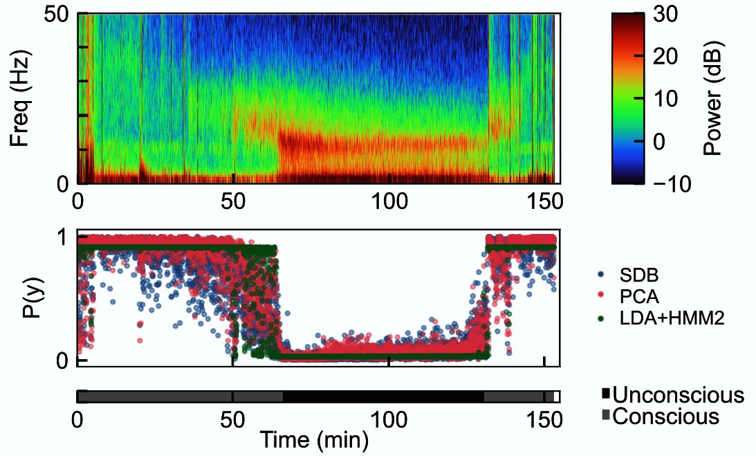
Over the last several years, Brown has joined forces with Picower Professor Earl K. Miller in these studies. Last year in Cell Reports, their labs showed that even among different anesthetic drugs, with their varying molecular mechanisms of action, they all have the same effect at the level of brain waves—they throw them out of phase with each other within the same brain hemisphere, thereby disrupting communication across the brain’s surface, or cortex. That turns out to be an apparently universal signature of unconsciousness (Miller has also shown this by a different measure working with MIT colleague Ila Fiete).
Brain waves are such a clear and powerful biomarker of unconsciousness under anesthesia that Brown and Miller have been able to devise a closed-loop system that can read out brainwaves and then automatically adjust dosing to maintain a target level of consciousness. They hope to someday deploy such systems in operating rooms to assist anesthesiologists.
Brain Imaging
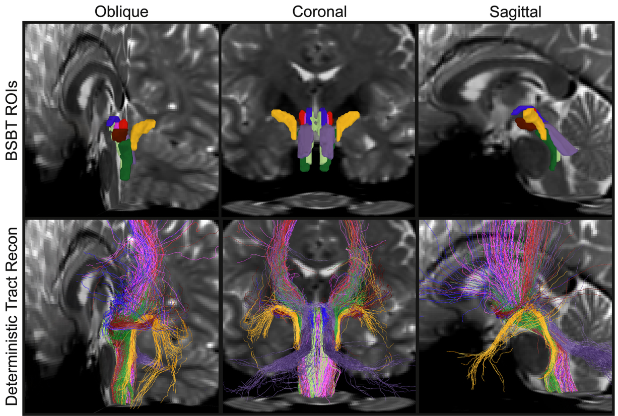
Brown’s lab also broke new ground last year by showing that imaging of the brainstem could deliver biomarkers for a variety of neurological conditions. The brainstem’s many bundles of nerve fibers help to govern essential functions such as consciousness, sleep, breathing, heart rate and motion. Because those bundles are small and tightly packed together, they’ve been very hard to image. But in a study in the Proceedings of the National Academy of Sciences, then graduate student Mark Olchanyi showed how he used AI to distinctly resolve eight brainstem bundles from live diffusion MRI images. Moreover, he demonstrated that retrospective analyses of images from patients using his “Brainstem Bundle Tool” could identify potential biomarkers of Parkinson’s disease, multiple sclerosis and traumatic brain injury. He even showed how the system could track the internal healing of a coma patient. The improvements visible in the imaging tool correlated with the improvement that enabled the patient to wake up after 7 months.
In another imaging advance, Sur and Osaki published a study last year in Light: Science & Applications that could yield another potential biomarker. With MIT Mechanical Engineering colleagues, the team invented a new microscope system that can peer especially deep into living brain tissue. The “photoacoustic” scope, which sends in light, but measures sound coming out, was able to detect NAD(P)H, a molecule tightly associated with cell metabolism in general and electrical activity in neurons in particular. Levels of the molecule are known to vary in conditions such as Alzheimer’s disease, Rett syndrome, and seizures, making it a potentially valuable biomarker.
The microscope needs more development before it could be used to image live human brains (for instance during surgeries), but like the other innovations above, it shows that, with research, scientists are finding many ways to help clinicians go beyond subjective symptoms. Biomarkers will increasingly make a mark on neurological and psychiatric care.


