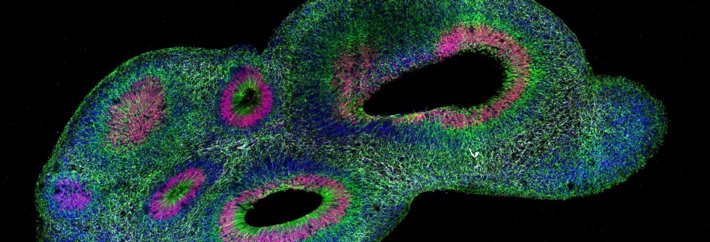
The ability to culture cerebral organoids, or “minibrains,” using stem cells derived from people has given scientists experimentally manipulable models of human neurological development and disease, but not without confounding challenges. No two organoids are alike and none of them resemble actual brains. To help researchers make meaningful scientific comparisons despite this variability, MIT neuroscientists and engineers developed a new pipeline for clearing, labeling, 3D imaging, and rigorously analyzing organoids called “SCOUT.”
SCOUT, described in 2020 in Scientific Reports, makes organoids optically transparent so they can be imaged with their 3D structure intact. The cleared organoids are then infused with antibody labels targeting specific proteins to highlight cellular identity and activity. The organoids are then imaged with a light-sheet microscope to gather a 3D picture of the whole organoid at single-cell resolution. Each organoid produces about 150 GB of data for automated analysis by SCOUT’s freely available software, principally coded by co-lead author Justin Swaney.
The software is able to detect every distinctly labeled cell and to recognize and track architectural patterns formed by the cells as the organoids develop or are affected in models of disease. Ultimately, the researchers were able to build a set of nearly 300 features on which organoids could be compared, ranging from the single-cell to whole-tissue level.

