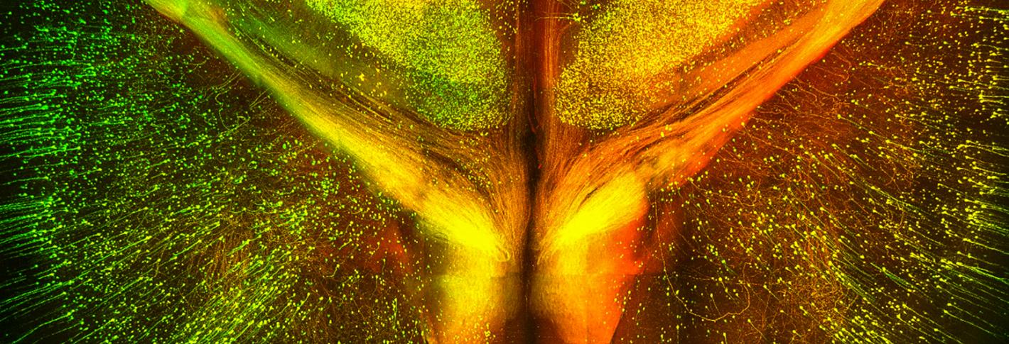
CLARITY makes tissue, like the brain, completely see-through. With a clear brain and additional biological techniques, an exceptionally detailed map of neuronal pathways can be made—inaccessible even from our best technologies a few years ago.
With this enhanced resolution of brain anatomy, neuroscientists are now able to look for the microscopic differences between healthy and dysfunctional brains. CLARITY further weds basic research to clinical practice, providing new views on biopsied and post-mortem human samples.
In 2015, a new shared CLARITY imaging core equipment facility was created to allow the Picower Institute to lead brain mapping microscopy methods to strategically make new advances and delve into unexplored areas of neuroscience research.
The facility includes hardware and software infrastructure for CLARITY technology. The equipment includes a high-content rapid throughput imaging microscope system from Leica Microsystems and Leica supporting software. The equipment is available to all Picower labs for use 24 hours per day, 7 days a week.
Videos and data collected using this new technology are shown at Brain Lunch, Plastic Lunch, the winter brain conference, and will also be shared at upcoming Gordon Conferences and Society of Neuroscience conference. Most notably are videos depicting clarified mouse and human patient post-mortem brains showing new pathological information for diseases such as Alzheimer’s.


